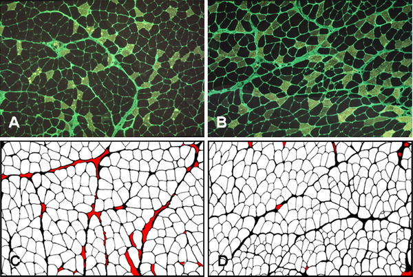
Fig. 2.

Immunohistochemical localization of fast myosin and fibronectin in soleus muscles from control (a) and trained (b) rats. Fibronectin staining was used for image analysis of muscles; the medial gastrocnemius muscles are illustrated from control (c) and trained (d) rats. Red areas represent areas not included in the measurements due to tears in the tissue or the presence of large blood vessels. Primary magnification 25×
