Fig. 4.

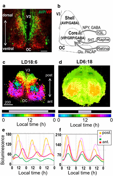
Photoperiodic clock in the SCN. a A coronal section of a mouse SCN immunolabeled with anti-AVP (green) and anti-VIP (red). Scale bar: 200 μm; OC, optic chiasm; V3, third ventricle. b Schematic drawing showing the cytoarchitecture of the SCN with major afferents and their neurotransmitters. Glu, glutamate; NPY, neuropeptide Y. c, d Phase maps for Per1-luc rhythms of horizontal SCN slices from mice housed under light–dark (LD) cycles 18:6 (L:3:00–21:00) (c) and 6:18 (L:9:00–15:00) (d). Pseudocolored bioluminescent images of peak phases of Per1-luc rhythms (each pixel = 3.7 × 3.7 μm). The peak phases are widely distributed in LD18:6 (from approximately 3:00 in the central SCN to 22:00 in the anterior SCN), and are consolidated between 9:00–14:00 in LD6:18. (see Ref. [116]). e, f, Bioluminescence from single pixels of horizontal SCN slices indicate a large regional phase distribution from the posterior (post) to anterior (ant) SCN in LD18:6, but are synchronized across the SCN as a whole in LD18:6 (see Ref. [93])
