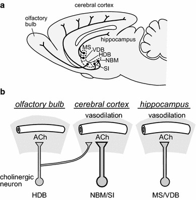
Fig. 5.

Summary of the basal forebrain cholinergic vasodilative system. a Diagram showing the cholinergic neurons in the basal forebrain and their fiber projections (modified from Sato et al. [10] based on Rye et al. [12]). b Comparative schema of ACh release and vasodilation in the three cholinergic pathways of the HDB-olfactory bulb, NBM/SI-cerebral cortex, and MS/VDB-hippocampus. HDB horizontal limb of the diagonal band of Broca, NBM nucleus basalis of Meynert; SI substantia innominate; MS medial septal nucleus; VDB vertical limb of the diagonal band of Broca
