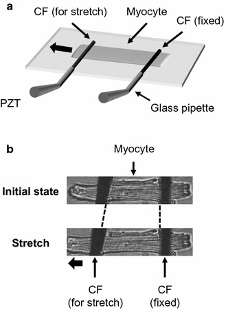
Fig. 1.

Overview of the stretch control system used to apply an axial stretch to the cell. a Both cell ends were held by a pair of carbon fibers (CF). The left CF was mounted on piezoelectric translators (PZT). b The myocyte was stretched from the initial state by moving the left CF with the PZT
