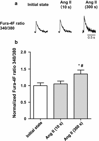
Fig. 3.

Change in [Ca2+] transient in cardiomyocytes from C57BL/6 J mice in the angiotensin II (Ang II) protocol. a Representative traces of Fura-4F fluorescence signals. After recording the initial state in normal Tyrode’s (NT) solution, the application of Ang II (1 μM) for 10 s left the [Ca2+] transient unchanged. However, application of Ang II for 300 s increased the [Ca2+] transient, similar to the SSC. b Summarized data for the change in [Ca2+] transient by Ang II application (n = 8). # P < 0.05 vs initial state; *P < 0.05 vs Ang II (10 s)
