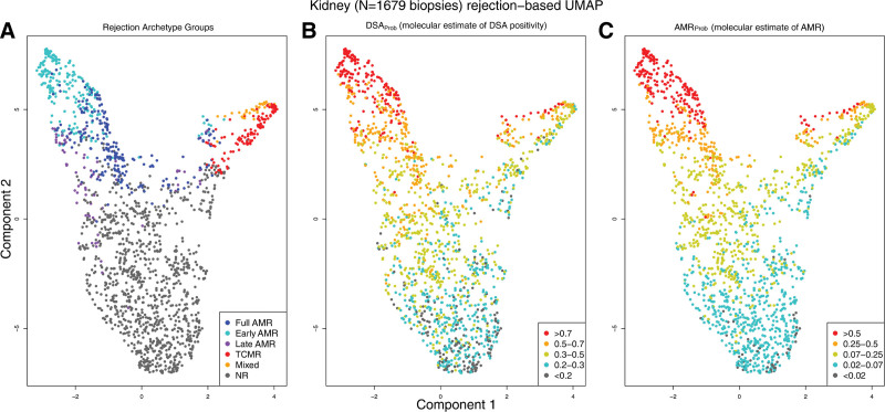
FIGURE 12.
UMAP projections of 1679 biopsies.63 All 1679 indication kidney transplant biopsy specimens, shown using UMAP, colored by (A) assigned rejection-based archetypal class, (B) increasing DSAProb classifier score, and (C) increasing AMRProb score. Biopsy samples with low probability of molecular rejection are located toward the bottom of Component 2 in all panels. Biopsy samples with rejection are located toward the upper region of Component 2, with AMR on the left and TCMR on the right of Component 1. AMR, antibody-mediated rejection; DSA, donor-specific antibody; TCMR, T cell–mediated rejection.

