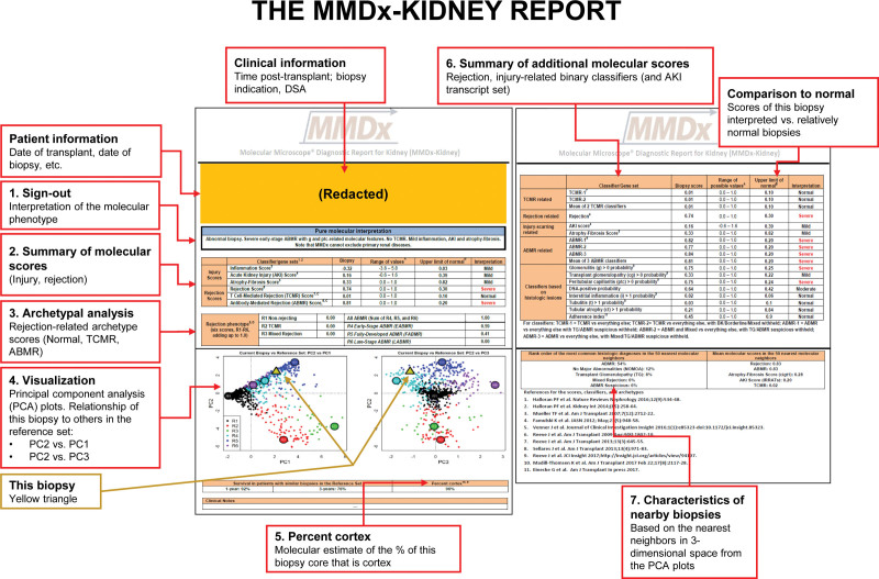
FIGURE 8.
The MMDx-Kidney report. Numbered items on the report are as follows: (1) The biopsy results are summarized by an expert reader, with comments on unusual features. This remains a necessary step because some biopsies have multiple or ambiguous features or rare phenotypes. (2) The molecular scores are summarized for the inflammatory disturbance, AKI (IRRAT), atrophy-fibrosis (the ci-classifier), and the all-rejection, TCMR, and AMR classifiers. (3) The archetype scores are summarized. Archetype scores are proportions, unlike binary classifier scores. Thus, a biopsy assigned to a particular archetype rejection cluster can have nearly as high a score in a second archetype but is only assigned to 1 group based on its highest (“dominant”) archetype score. (4) The rejection classifier scores are used to locate the position of the new biopsy (triangle) in relationship to the biopsies in the locked, N = 1208 reference set, which are colored by their rejection archetype states and shown in PCA plots: PC2 versus PC1 (left panel) and PC2 versus PC3 (right panel). (5) The percent cortex is estimated by NPHS2 (podocin) expression. Low %cortex (<10%) can affect some scores, eg, inflammation, cg-classifier, and late-stage AMR. (6) Details of selected rejection and injury scores of interest are presented and compared to relatively normal biopsies. (7) The characteristics of this biopsy are compared to its nearest neighbors in the reference set. AKI, acute kidney injury; AMR, antibody-mediated rejection; cg, transplant glomerulopathy; IRRAT, injury- and rejection-associated transcript; MMDx, Molecular Microscope Diagnostic System; PCA, principal component analysis; TCMR, T cell–mediated rejection.

