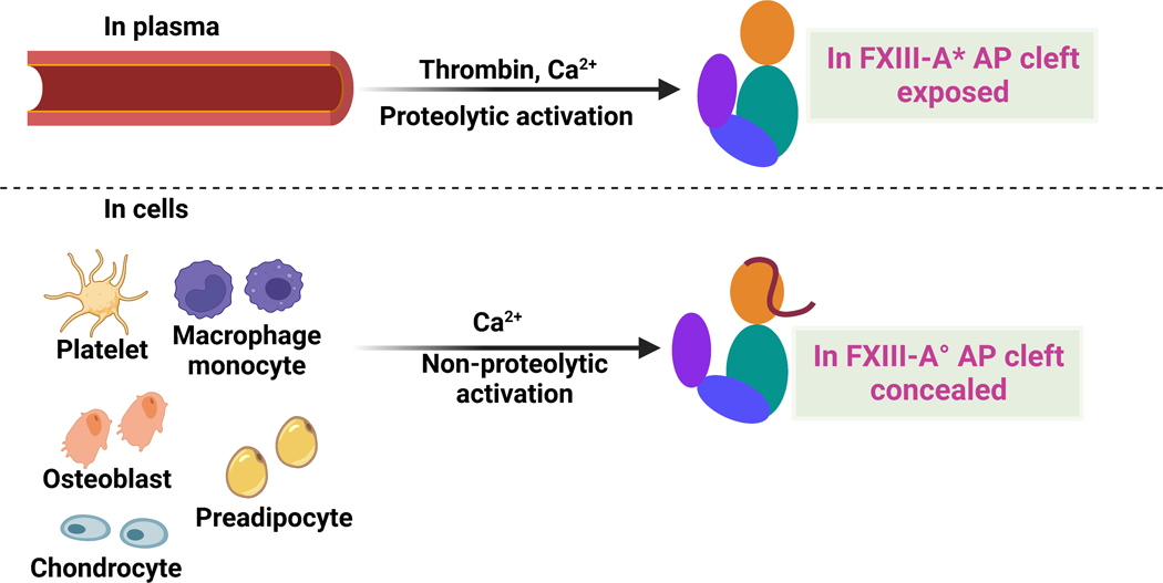
Fig. 1. Cartoon models highlighting FXIII locations and activation strategies.
pFXIII undergoes proteolytic activation by thrombin to form active FXIII-A* in which AP is removed and AP cleft is exposed. cFXIII is expressed in a variety of cells including platelets, monocytes, macrophages, chondrocytes, osteoblasts and preadipocytes. cFXIII undergoes nonproteolytic activation to form FXIII-A° in which AP is still associated with A subunit. In resting platelets and monocytes, FXIII is of cytoplasmic localization, however upon activation of these cells, FXIII-A is exposed on the surface of these cells. Cartoon model is created with BioRender.com.

