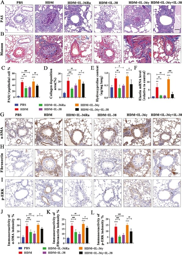
Figure 7.

IL-38 or blocking IL-36R alleviated airway remodeling and the expression of p-ERK1/2 in an HDM-induced asthma murine model. (A and B) Representative images of PAS staining and Masson staining in lung tissues from each group. Scale bar = 50 μm (×200). (C and D) Quantification of PAS staining and Masson staining in each group. (E) The HYP content in lung of each group mice was measured by HYP assay kit. (F) Quantification of elastin mRNA level in the mice lung tissues of each group. (G–I) Representative immunoreactivity images of α-SMA, Fibronectin and p-ERK1/2 in lung tissues from each group. Scale bar = 50 μm (×200). (J–L) Quantification of the expression of α-SMA, Fibronectin , and p-ERK1/2 in each group. Bar diagrams and data are presented as the mean ± standard deviation (SD). * vs. PBS group; # vs. the indicated group. *,#P < 0.05; **,##P < 0.01; ***,###P < 0.001
