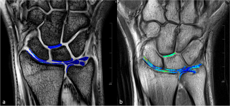
Fig. 2.
Three-Tesla images of the first echo of the T2*-weighted multi-echo gradient-echo sequence (a) and T2-weighted multi-echo spin-echo sequence (b) of a healthy volunteer with superimposed color-coded maps of T2* and T2 values. Regions of interest (ROIs) were placed in the cartilage of the distal radius, in the proximal scaphoid, in the proximal radial and ulnar lunate, in the distal radioulnar joint, and in between the distal lunate and proximal capitate. Three ROIs were placed at the triangular fibrocartilage complex: at the central disk and at both the foveal and apical attachments. The same analysis was performed for 7-T MRI

