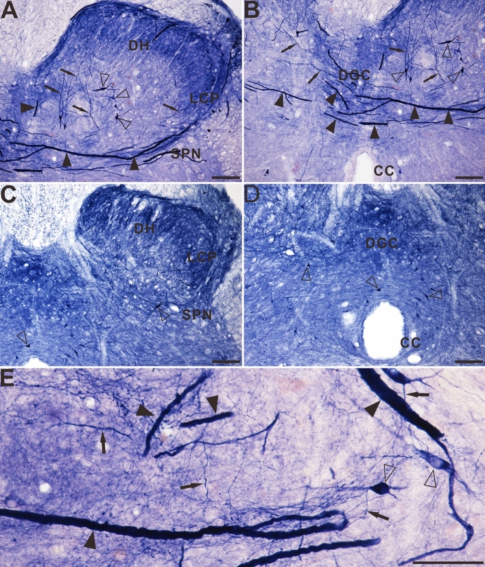
Figure 1.
Microphotographs of NADPH-d positive reactivity in aged and young dogs at the sacral (S2 segment) spinal cord. All of the transverse sections are taken at the same levels. Note intense and abnormal NADPH-d positive megaloneurites (black arrowheads) in the LCP (A) and DGC (B) in the sacral segment of aged dogs compared with young dogs (C) and (D). The NADPH-d positive megaloneurites in the sacral spinal cord of aged dogs are completely different from the surrounding normal fibers and neurons (E). Open arrowheads: NADPH-d positive neurons, black arrows: normal NADPH-d positive neurites, black arrowheads: megaloneurites. Scale bar in (A)–(D) = 100 μm, in (E) = 50 μm.

