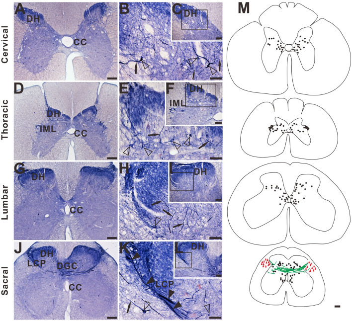
Figure 2.
Microphotographs showing the distribution of NADPH-d staining in transverse sections of the aged dog spinal cord at different segmental levels. Few neurons (open arrowheads) and normal fibers (black arrows) staining in the dorsal horn (DH) is present in cervical (A), thoracic (D) and lumbar (G) segments. Intense and abnormal fiber (black arrowheads) staining in the dorsal gray commissural (DGC) and LCP is present in the sacral (J) segment. (B), (E), (H), and (K) show higher magnifications from insert (C), (F), (I), and (L), respectively. (M) Schematic diagram of NADPH-d activity taken from the cervical, thoracic, lumbar, and sacral spinal cord segments. Neuronal cell bodies are indicated as filled circles (black) on both sides of each figure. Each filled circle represents one NADPH-d positive neuron. Dense NADPH-d stained megaloneurites are represented by cords (green). The triangle symbols (red) indicate NADPH-d activity in the white matter. NADPH-d stained neurons and fibers from 5 sections are plotted on a drawing of the transverse section of the aged dogs of the spinal cord at indicated segmental levels. Open arrowheads: NADPH-d neurons, black arrows: normal NADPH-d positive neurites, black arrowheads: megaloneurites. Scale bar in (A), (D), (G), (J), (M) = 200 μm; (C), (F), (I), (L) = 100 μm; (B), (E), (H), (K) = 50 μm.

