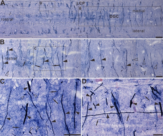
Figure 7.
The Megaloneurites in horizontal sections in the DGC in the sacral segment of aged dogs. (A) Horizontal sections in the low power microphotograph confirmed that the megaloneurites are organized in a regular interval vertical to the rostrocaudal axis. (B) is the magnification of (A). (C), (D) High power microphotographs demonstrated a significant difference between the megaloneurites and normal fibers and neurons. Open arrowheads: NADPH-d neurons, black arrows: normal NADPH-d neurites, black arrowheads: abnormal megaloneurites. Scale bar in (A) = 200 μm, in (B) = 100 μm, in (C), (D) = 50 μm.

