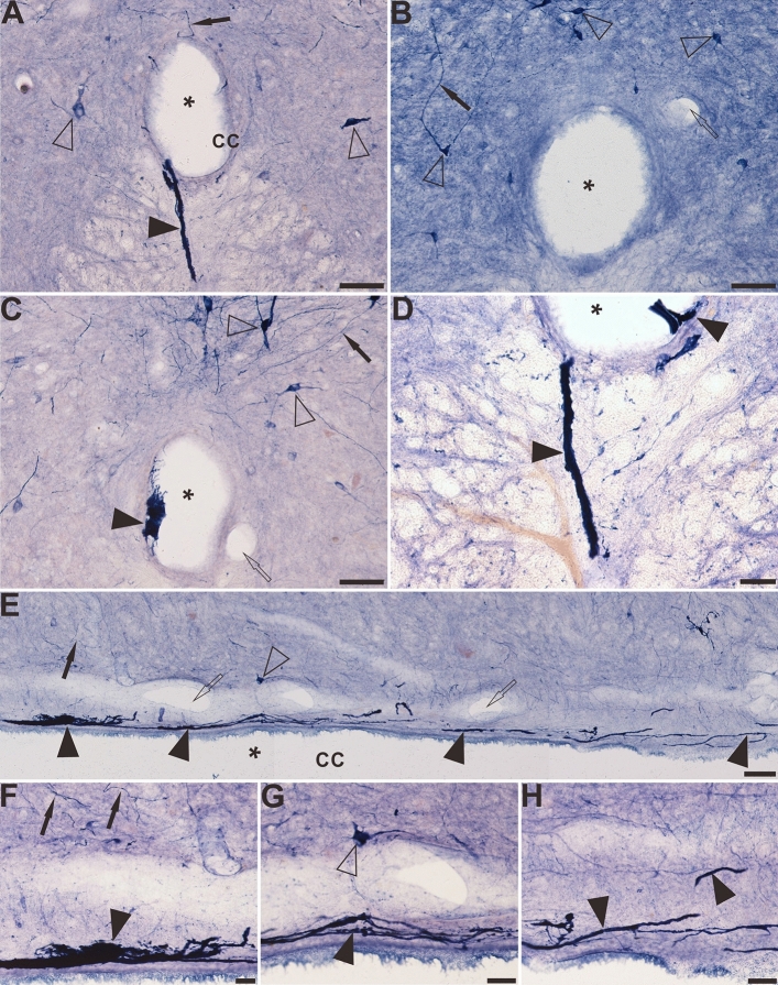
Figure 8.
The distribution of NADPH-d positive abnormality in the sacral-coccygeal spinal cord of aged dogs. (A), (C) and (D) demonstrate the location and morphology of the NADPH-d abnormalities on the transverse section of aged dogs. Some positive fibers and structures cross the ependymal cells and reach the lumen of the CC (A). There are no aberrant structures in the CC of young dogs (B). Note abnormal mass-like or strand-like NADPH-d positive structures (black arrowheads) around the CC of aged dogs. Both intra-CC and extra-CC alterations are detected in (D). Megaloneurites indicated ventrally bridging from the CC to the anterior median fissure (A) and (D). (E)–(H) shows the location and morphology of the NADPH-d abnormalities along the CC in the horizontal section of aged dogs. Open arrowheads: NADPH-d positive neurons, black arrowheads: NADPH-d positive abnormalities, open arrows: the vascular structures, black arrows: normal NADPH-d positive neurites, the asterisk indicates lumen of CC. Scale bar in (A)–(C), (E) = 50 μm, in (D), (F)–(H) = 20 μm.

