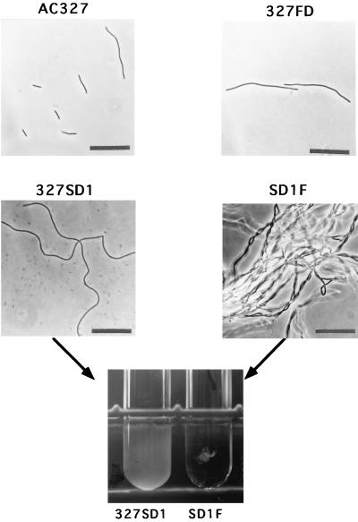
FIG. 6.
Phase-contrast microscopy of AC327 (wild-type), 327FD (cwlF), 327SD1 (sigD), and SD1F (cwlF sigD) cells and a picture of the test tube cultures of 327SD1 and SD1F. The pictures of AC327, 327FD, 327SD1, and SD1F were taken at OD600s of 0.263, 0.117, 0.146, and 0.205, respectively. Bar, 25 μm. To measure the OD600 of the SD1F strain, a small amount of lysozyme was added to the samples just before vigorous vortexing.

