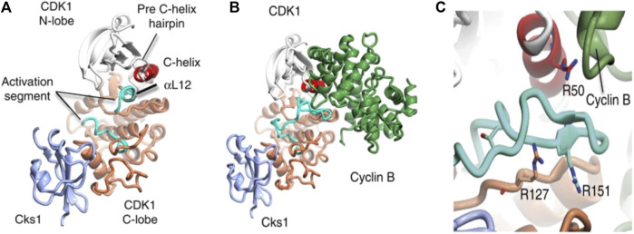
FIGURE 1.
Structure of CDKI binding to CKs1 and complexation with cyclin B. The complexes shown above depict (A) CDK1-Cks1 (B) CDK1-cyciln B-Cks1 (C) residues of CDK1. In each image, the following colours apply; CDKI N-lobe (white), CDK1 C- lobe (coral), Cks subunit (ice-blue), C-helix (red), activation segment (cyan), cyclin subunit (lawn green) (Brown et al., 2015).

