Fig. 3.
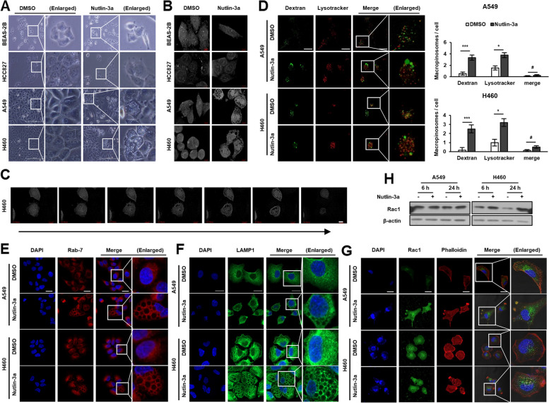
Nutlin-3a induces huge cytoplasmic vacuoles in KRAS MT/p53 WT NSCLC cells. Cells were treated with nutlin-3a (30 μM) for 6 (D, H) or 24 h (A, B, E–H). A, B Representative live cell images captured by light (A) and holotomography microscope (B) show huge cytoplasmic vacuoles in KRAS MT/p53 WT but not KRAS WT cells. C Time-lapse video capture images (24 h) of H460 obtained by holotomography microscopy show increase in macropinosomes volume with time and membrane rupturing in the end. D Nutlin-3a induced formation of huge vacuoles were incorporated with dextran (green), but not merged with LysoTracker (red). Representative live images were obtained by confocal laser scanning microscopy (left). Number of macropinosomes were quantitated from the captured image (right) (n ≥ 8). E–G Representative images obtained by confocal laser scanning microscopy. Fixed cells were stained with Rab7 (E), LAMP1 (F), or Rac1 (green), and rhodamine–phalloidin (red) (G) and were counterstained with DAPI (blue). H Western blotting of Rac1 expression. *p < 0.05, **p < 0.01, ***p < 0.001 compared with control. Scale bar: 200 μm (A), 7 μm (B, C), and 20 μm (D-G)

