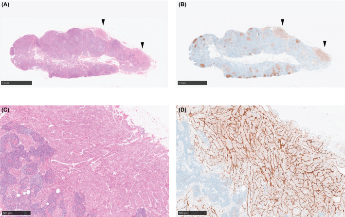
FIGURE 1.

Nodal lymphangioleiomyomatosis (LAM) in a common iliac lymph node. (A, C) Hematoxylin and eosin (HE) staining showed the bundles of spindle cells and nests of epithelioid cells in a common iliac lymph node. (B, D) D2‐40 (podoplanin) expression in the lymphatic endothelium lining the spindle‐cell bundles and epithelioid‐cell nests. (A, B) Loupe images capturing the whole lymph node. Representative LAM lesions are shown with black arrowheads. Scale bar: 5 mm. (C, D) Representative magnified views. Scale bar: 500 μm.
