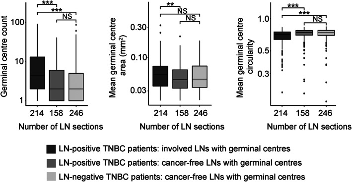
Figure 4.

Properties of smuLymphNet‐captured GCs. The LN sections were separated into (1) involved LNs with GCs from LN‐positive TNBC patients, (2) cancer‐free LNs with GCs from LN‐positive TNBC patients, and (3) cancer‐free LNs with GCs from LN‐negative TNBC patients. Left to right: boxplots display distribution per LN section for GC count, mean GC area (mm2), and mean GC circularity. Statistical significance was assessed using a two‐sided Wilcoxon rank sum test (**p ≤ 0.01, ***p ≤ 0.001, NS = not significant).
