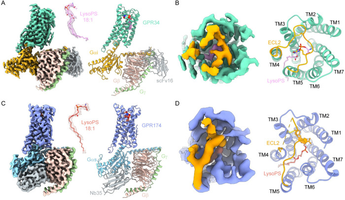
Fig 1. Overall structures of GPR34-Gi complex and GPR174-Gs complex.
(A) Cryo-EM density map and model of GPR34-Gi complex. Density of LysoPS is shown as mesh in the middle and colored plum. (B) The map and model of ECL2 of GPR34-Gi complex (extracellular view). (C) Cryo-EM density map and model of GPR174-Gs complex. Density of LysoPS is shown as mesh in the middle and colored salmon. (D) The map and model of ECL2 of GPR174-Gs complex (extracellular view). GPR34 is colored marine green. GPR174 is colored blue. ECL2 is colored orange. Gαi, Gαs, Gβ, and Gγ subunits are colored gold, cyan, pink, and light green, respectively. ScFv16 and Nb35 are colored grey.

