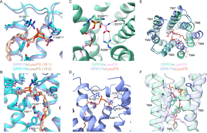
Fig 3. Comparison of LysoPS recognition by GPR34 and GPR174.
(A) Interactions between GPR174 and the polar head of LysoPS (18:0) or LysoPS (18:1). (B) Interactions between GPR174 and the acyl tail of LysoPS (18:0) or LysoPS (18:1). The double bond of LysoPS (18:1) is colored red; the corresponding single bond in LysoPS (18:0) is colored orange. (C) Charged interactions between LysoPS and GPR34 in the positively charged pocket. (D) Charged interactions between LysoPS and GPR174 in the positively charged pocket. (E) Structural superposition of LysoPS bound GPR34 and GPR174 (extracellular view). (F) Comparison between acyl tails of LysoPS binding in GPR34 and GPR174. Polar or charged interactions are depicted as black dashed lines.

