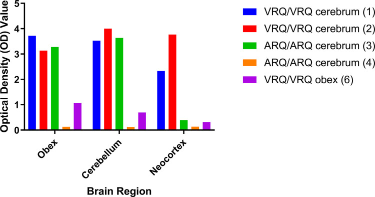
Fig 3. Quantitative analysis of PrPSc present in brainstem, cerebellum, and neocortex.
Differences in the amount of PrPSc present in brain regions observed by IHC (Fig 2) can be assessed using EIA. The VRQ/VRQ sheep (sheep 1 and 2) have high OD values in all three brain regions. The positive ARQ/ARQ sheep (sheep 3) had high OD values for brainstem and cerebellum but a lower score for the neocortex compared to VRQ sheep that received the same inoculum. The ARQ/ARQ sheep (sheep 4) without immunoreactivity by IHC also was negative by EIA in all three brain regions. A VRQ/VRQ sheep challenged with the scrapie agent from deer brainstem (sheep 6) was positive by EIA but with low OD values relative to sheep challenged with inoculum from deer cerebrum.

