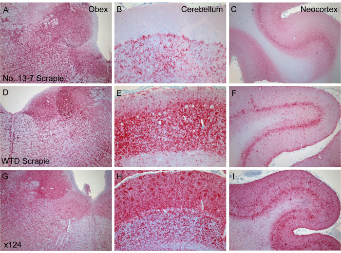
Fig 8. Comparison of PrPSc accumulation between scrapie strains.
PrPSc distribution differences are noticeable in various brain regions of sheep inoculated with different scrapie isolates. The original No.13-7 scrapie inoculum in sheep is represented in panels A-C. WTD scrapie in sheep, sheep 2, has more PrPSc labeling in the cerebellum (E) and neocortex (F) compared to No.13-7 (B, C) and also similar, but less, labeling to x124 (G-I).

