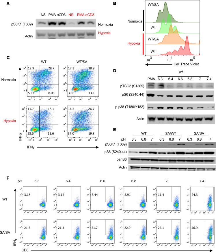
Figure 5. SA mutation in CD8+ T cells amplifies mTORC1 activation under cellular stress.
(A) Resting CD8+ T cells stimulated with PMA or TCR (αCD3) in normoxic or hypoxic (2% O2) conditions for 15 minutes and assayed by immunoblot for mTORC1 activity via pS6K1. (B) Proliferation analysis of stimulated OTI CD8+ T cells with WT or WT/SA TSC2 in normoxia or hypoxia. Cells expressing WT/SA proliferate more in both conditions. (C) Flow cytometry analysis for cytokine function from IL-2 generated cytotoxic WT versus TSC2-SA mutant CD8+ T cells that were rechallenged overnight in normoxic or hypoxic (2% O2) conditions. (D) Immumoblot for phosphorylated TSC2-S1365, S6, and p38 MAP kinase from IL-2 pretreated CD8+ T cells and then cultured in neutral or more acidic media. (E) Activated WT, mutant TSC2 WT/SA and SA/SA CD8+ T cells were exposed to various media at various pH for 90 minutes and assayed by immunoblot analysis for mTORC1 activity. (F) Effector TSC2 WT or TSC2 SA/SA CD8+ T cells were stimulated with PMA and ionomycin in various pH level media to assess IFN-γ via flow cytometry. Data are representative of at least 3 independent experiments, except E and F, with 2.

