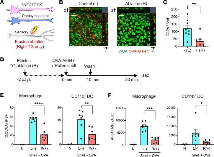
Figure 7. Trigeminal nerve ablation inhibits RW pollen shell–stimulated GAP formation and early antigen uptake.
(A) The trigeminal (TG) nerve was ablated by bipolar coagulation. (B and C) GAP formation 5 minutes after instillation of OVA-AF647 and pollen shells. Representative image (B) and quantitation (C) (n = 8). Scale bars: 50 μm. **P < 0.01 by 2-tailed, paired Student’s t test. (D) Experimental diagram of the early antigen passage after TG ablation. (E and F) The frequencies of OVA-AF647+ cells (E) and the mean fluorescence intensity (MFI) (F) of the indicated cell types (n = 2–7). The nontreated samples were used for setting the positive gate and were excluded from the statistical analysis. In C, E, and F, – or (–) indicates no TG ablation and + or (+) indicates TG ablation. *P < 0.05, **P < 0.01, ***P < 0.001, ****P < 0.0001 by 2-tailed, paired Student’s t test. B6 mice were used for all experiments.

