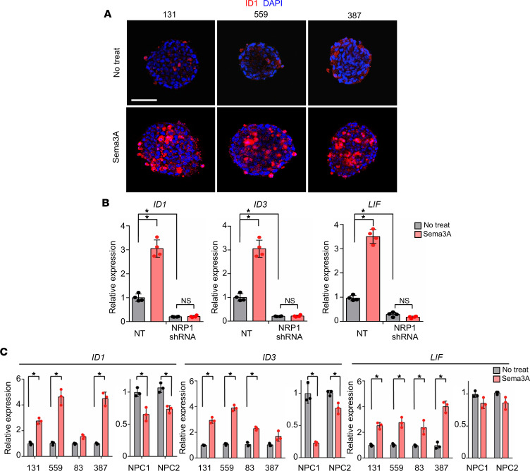
Figure 4. The Sema3A/NRP1 axis in GBM activates canonical TGF-β signaling.
(A) Immunostaining images of ID1 in 131, 559, and 387 GBM cells treated with rSema3A. ID1-positive cells are shown in red. Scale bar: 50 μm. (B) Levels of the representative TGF-β pathway genes (ID1, ID3, and LIF) in the NT or NRP1-KD 131 GBM cells. n = 4. (C) Levels of ID1, ID3, and LIF mRNAs in GBM cells and NPCs treated with rSema3A. n = 3. Data represent mean ± SD. *P < 0.01 by 1-way ANOVA with Tukey’s multiple comparison test in B and C.

