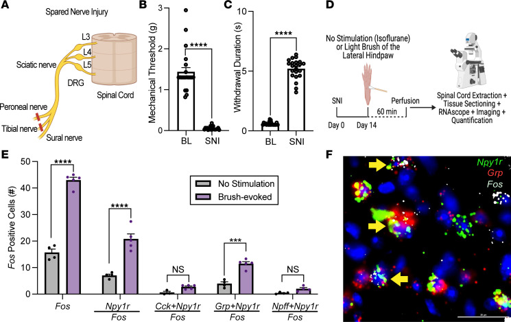
Figure 4. Non-noxious mechanical stimulation increases neuronal activity in Grp/Npy1r-INs.
(A) Schematic representation of the spared nerve injury (SNI) model of neuropathic pain. (B and C) SNI produces mechanical (****P < 0.0001) and cold hypersensitivity (****P < 0.0001) in the ipsilateral hind paw when assessed 14 days after injury. Unpaired 2-tailed t test (n = 24 mice/group). (D) Experimental timeline for SNI, light brush of the lateral hind paw, and FISH labeling for Fos in Y1-IN subpopulations in the lumbar dorsal horn of SNI mice. (E) Light brush increases Fos-positive cells in the superficial dorsal horn (****P < 0.0001) primarily in Npy1r neurons (****P < 0.0001) and Grp/Npy1r neurons (***P = 0.0001) but not Cck/Npy1r neurons (P = 0.7371) or Npff/Npy1r neurons (P = 0.8095) (2-way ANOVA: mRNA expression × stimulation, F4, 33 = 1.215, P < 0.0001, Holm-Šídák post hoc tests) (n = 3–5 mice/group). Each data point indicates the average of 2–4 quantified sections/mouse. (F) Representative example of Grp/Npy1r-INs coexpressing light brush–evoked Fos. Scale bars: 25 μm. Yellow arrows indicate colocalization. Data shown as mean ± SEM.

