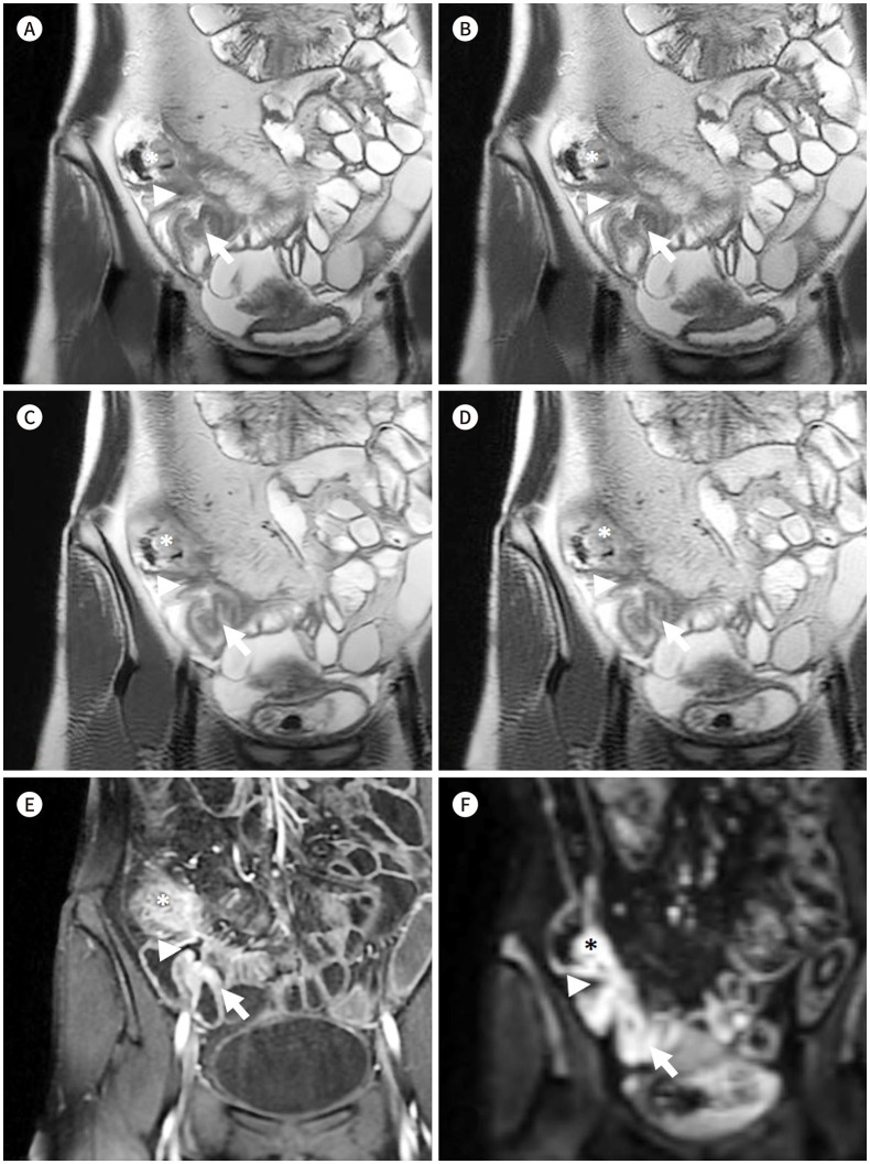
Fig. 3. Coronal SSFSE T2-weighted images of a 30-year-old male patient with Crohn’s disease.
A-D. MBH-DLR (A), MBH-CR (B), SBH-DLR (C), and SBH-CR (D), respectively. The DLR images (A, C) show reduced noise with sharper margin of bowel wall compared with those in the CR images (B, D); better delineation of the bowel wall and of the inflammation (asterisks) in the cecum and terminal ileum with an enteroenteric fistula (arrows) and an enterocolic fistula (arrowheads); moreover, both readers gave all the four sequences the same scores regarding mural thickness, mural signal intensity, and perimural signal intensity.
E, F. Contrast-enhanced T1-weighted image (E) and diffusion restriction image (F) (b = 900) of the corresponding area. Contrast enhanced image show mural hyper-enhancement and wall thickening of cecum and terminal ileum (asterisks), enteroenteric fistula (arrows) and enterocolic fistula (arrowheads) which show diffusion restriction, suggesting active inflammation.
CR = conventional reconstruction, DLR = deep-learning based reconstruction, MBH = multiple-breath-hold, SBH = single-breath-hold

