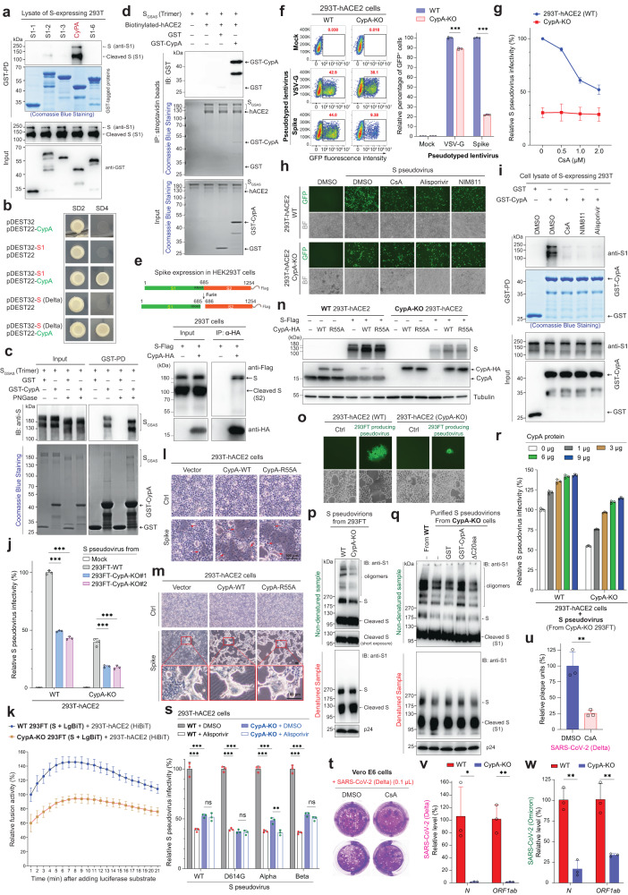
Fig. 1.
Host CypA facilitates SARS-CoV-2 infection by interacting with and regulating spike on the virions. a SARS-CoV-2 spike was found to interact with human CypA. 293 T cells expressing S were lysed in Co-IP buffer and subjected to a GST pull-down (PD) assay with recombinant GST-CypA. Samples were analyzed by anti-GST or anti-S1 immunoblotting (IB). b S1 or S binds to human CypA in the Y2H system. Plasmids that express S1 (or S) and/or CypA were transfected to yeast cells, and the growth of yeast on SD2 and SD4 agar plates was estimated after about two weeks. c Trimeric SGSAS protein interacts with CypA in vitro. Non-cleaved SGSAS mutant (substitution of RRAR (682-685aa) by GSAS) was purified from 293 T primarily in trimeric forms. SGSAS trimers was subjected to GST- or GST-CypA-based PD assay supplemented with or without PNGase, at 4 °C overnight. d S-hACE2 complex can recruit CypA. Trimeric SGSAS was incubated with biotinylated hACE2 and GST or GST-CypA, and then Co-IP with streptavidin beads was conducted. e CypA prefers full-length S, but not its cleaved form (S2). 293 T cells transfected with S and CypA-HA plasmids were lysed in Co-IP buffer and subjected to anti-HA co-IP and subsequential western blots. f CypA deficiency robustly blocks S pseudovirus infection. WT or CypA-KO 293T-hACE2 cells were incubated with S pseudovirus or VSV-G pseudovirus. After 24 h, cells were analyzed by flow cytometry. Shown in the left panel are flow cytometric plots with the proportions of GFP-positive cells. A comparison of percentages of GFP-positive cells are present in the right panel (n = 3). g Removal of CypA in targeted cells eliminates the inhibitory effect of CsA on viral infection. WT or CypA-KO 293T-hACE2 was infected by S pseudovirus with different concentrations of CsA. GFP-positive cells were detected by flow cytometry and relative infectivity was calculated (n = 3). h CsA and its derivatives inhibit S pseudovirus infection by targeting CypA. WT or CypA-KO 293T-hACE2 cells were incubated with equal virions and indicated inhibitors (1 μM) for 24 h. GFP-fluorescent images and bright field (BF) images were taken by microscopy. Scale bars, 50 μm. i CsA and its derivatives block S recruitment to CypA. Cell lysates of S-expressing 293 T cells, GSH beads, GST-CypA, and CypA inhibitors (with final 1 μM) were incubated in PD buffer for 2 h. GSH beads were then collected, washed, and analyzed by Coomassie blue staining of SDS-PAGE gel or anti-S1 IB. j Deficiency of CypA in packaging cells impairs viral infectivity. WT or CypA-KO 293T-hACE2 cells were infected by viruses produced from one WT or two CypA-KO 293FT packaging cell lines for 24 h. Cells were analyzed by flow cytometry (n = 3). k CypA depletion in S-expressing cells attenuates S/hACE2-mediated cell–cell fusion. 293T-hACE2 cells expressing HiBiT were incubated with WT or CypA-KO 293FT cells expressing S and LgBiT. 5 h later, substrate furimazine was added to cells and luminescence intensities were detected (n = 3). l Overexpression of CypA exacerbates S-mediated cell-cell fusion in 293T-hACE2. Empty vector or S-expressing plasmid was co-transfected with the plasmid encoding WT or R55A CypA in 293T-hACE2 cells for 18 h. Cell fusion events were analyzed by microscopy. Shown are the bright field (BF) images (scale bars, 100 μm). Red arrowheads mark cell-cell fusion. m CypA dramatically promotes the formation of S-mediated giant syncytia in 293T-hACE2. Cells were transfected with S and CypA or its R55A mutant for about 30 h. BF images were taken by microscopy. Scale bars, 100 μm. n CypA expression in host cells stabilizes S protein. Plasmids expressing S and/or CypA-HA (or CypA-R55A-HA) were co-transfected into WT or CypA-KO 293T-hACE2 for 24 h. Samples were collected and subjected to anti-S1, anti-CypA, or anti-Tubulin IB analysis. o Cell fusion-mediated viral transmission in 293T-hACE2 is blocked by CypA deficiency. 293FT cells producing S pseudovirus were co-cultured with WT or CypA-KO 293T-hACE2 cells for 24 h, and then images were acquired by microscopy. Scale bars, 50 μm. p Deficiency of CypA in the packaging cells decreases S oligomers on the virions. S pseudovirus particles produced from WT or CypA-KO 293FT cells were lysed to prepare non-denatured and denatured samples for anti-S1 western blot. q CypA facilitates S oligomers formation on virions. Purified S pseudovirions from CypA-KO cells was incubated with GST, GST-CypA, or GST-CypA-△C20aa at 37 °C for one hour. Non-denatured and denatured samples of virions were analyzed by anti-S1 or anti-p24 western blot. r Incubation with recombinant CypA protein increases the infectivity of S pseudovirus. Purified S pseudovirions from CypA-KO cells was incubated with different amounts of recombinant CypA protein at 37 °C for one hour. These S pseudovirions were then utilized to infect WT or CypA-KO 293T-hACE2 for 24 h. Cells were analyzed by flow cytometry (n = 3). s Alisporivir inhibits the infection of SARS-CoV-2 variants (D614G, Alpha, or Beta) pseudovirus by targeting CypA. WT or CypA-KO 293T-hACE2 treated with or without alisporivir (1 μM) was infected by different pseudovirus and analyzed by flow cytometry (n = 3). t, u CsA strongly restricts plaque formation of SARS-CoV-2 Delta. Vero E6 cells treated with or without CsA were incubated with 0.1 μL SARS-CoV-2 Delta and seeded into the 12-well plates. When the plaques were formed, cells were fixed and stained with 0.5% (w/v) crystal violet. Shown in t are images, and shown in u is the quantitative data of plaque-forming units (n = 3). v, w Knockout of CypA in 293T-hACE2 cells blocks infection of SARS-CoV-2 Delta or Omicron. WT and CypA-KO 293T-hACE2 cells were infected by Delta (v) or omicron (w) variant. 48 h later, culture supernatant was subjected to qPCR analysis of viral gene N or ORF1ab. Data are presented as mean ± SD (Student’s t-test). *p < 0.05, **p < 0.01, ***p < 0.001, ns: no significance

