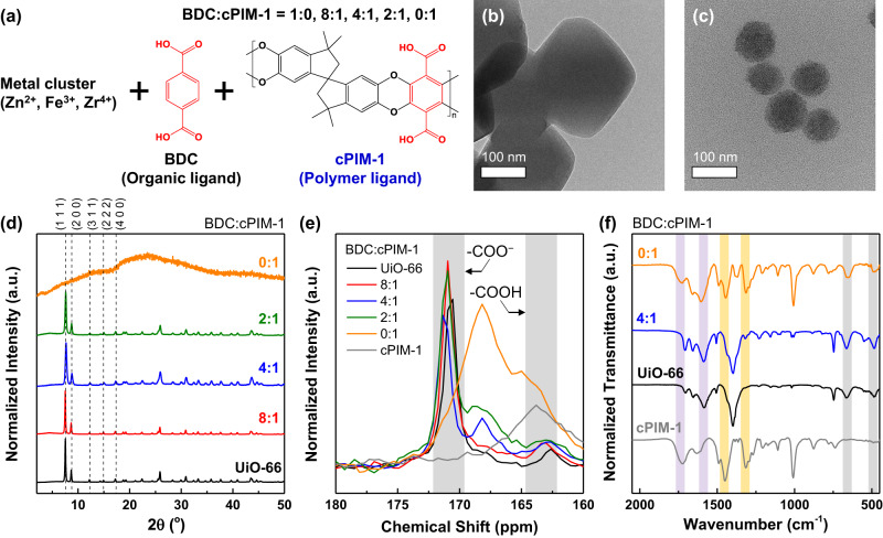
Fig. 2. Characterization of polyMOFs.
a Synthesis of polyMOF nanoparticles containing cPIM-1 ligand. Transmission electron microscopy (TEM) images of b UiO-66 and c polyUiO-66(4:1) nanoparticles. d Powder x-ray diffraction (XRD) patterns, e solid-state 13C nuclear magnetic resonance (NMR) spectra, and f Fourier-transform infrared spectroscopy (FT-IR) spectra of polyUiO-66 samples depending on BDC:cPIM-1 ratio used for their synthesis. Note: yellow highlights: methylene (CH2) stretching/bending vibration modes and the C−O stretching mode, purple highlights: carboxylate bands for cPIM-1 and the red-shifted peaks for polyUiO-66 samples, and gray highlights: Zr oxo clusters in polyUiO-66 samples.

