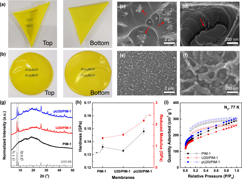
Fig. 5. Characterization of MMMs.
Photo images of a U20/PIM-1 and b pU20/PIM-1 membranes. Cross-sectional scanning electron microscopy (SEM) images of c, d U20/PIM-1 and e, f pU20/PIM-1 membranes. Red arrows indicate the significant agglomerates and interfacial voids in U20/PIM-1 mixed matrix membrane (MMM). g XRD spectra, h mechanical properties calculated from the nanoindentations tests (error bars represent standard deviations from 5 different analyses), and i N2 sorption isotherms at 77 K (filled symbols: adsorption and unfilled symbols: desorption).

