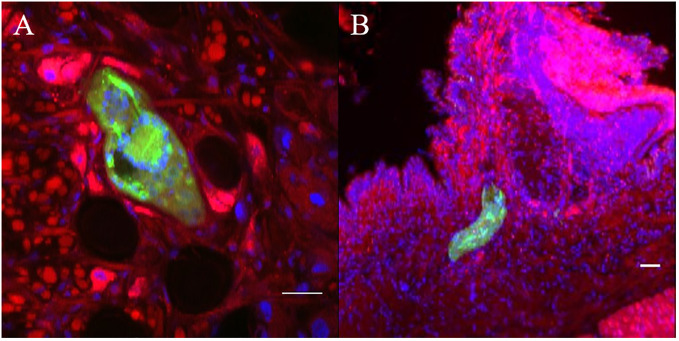
Figure 7.
Confocal imaging of Biomphalaria glabrata and Schistosoma mansoni through vibratome sections. (A, B) CMFDA-labeled sporocysts were highlighted in green, snail actin filaments were stained with phalloidin in red, and snail cell nuclei were counterstained with DAPI in blue. Scare bares indicates 20 μm.

