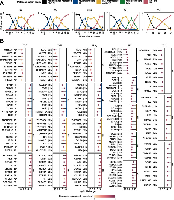
Fig. 3.
Temporal profiles obtained from NMF. A The pattern matrix for each CD4+ T-cell population from the Discovery Set is shown as continuous profiles, with samples assigned to time points of activation. We scaled each column in the pattern matrix to sum up to one. Dots depict median weights for all samples from identical analysis time points. Vertical lines represent interquartile ranges. We annotated and colored the metagenes based on their maximum median values across all analysis time points. The time point with the maximum median value is depicted in the legend. B Top 10 genes associated with metagenes for each T-cell population. For each CD4+ T-cell population and gene used for NMF, we used the highest absolute “confect” value estimated in the DGEA across all contrasts (e.g., 12 h vs. 0 h). Genes are ranked by “confect” values. Dots represent log2 fold changes for contrasts with the highest absolute “confect” value. The time point to the right of the gene represents the contrast with the highest absolute “confect” value. For example, “12 h” represents the following contrast: a sample group activated for 12 h compared to unactivated samples of the same group. The color of the dots corresponds to rank normalized average expression values of the activation group in contrast with the highest absolute “confect.” The inner end of the horizontal line shows the “confect” value (inner confidence bound). NFKBID denotes NF-κB

