FIG. 3.
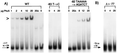
Gel mobility shift assays of PhcA binding to −117 to +36 xpsR promoter fragments with mutations in the PhcA binding site. (A) DNA fragments containing the −117 to +36 sequences of wild-type PxpsR (WT) or PxpsR with the indicated sequence alterations were isolated, labeled with [α-32P]dATP, and incubated with 0 to 20 μg of purified PhcA protein or with 20 μg of a similar protein preparation from E. coli lacking phcA (20Δ). Reaction mixtures were electrophoresed, and the dried gels were subjected to autoradiography as described previously (30); quantitative results were obtained by phosphorimaging and are summarized in Table 3. (B) A labeled PxpsR DNA fragment lacking sequences upstream of nucleotide −77 (Δ > −77) was analyzed as described for panel A. The symbol > to the left of the gel indicates the position of retarded (bound) fragment.

