Fig. 1.

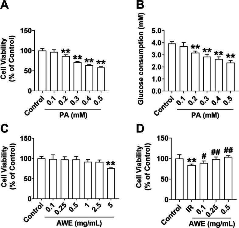
AWE increased the cell viability in IR HepG2 cells. A CCK8 assay was performed to examine the viability of HepG2 cells exposed to indicated concentrations of PA for 24 h. B Glucose consumption was measured in HepG2 cells exposed to indicated concentrations of PA for 24 h. C CCK8 assay was performed to examine the viability of HepG2 cells exposed to indicated concentrations of AWE for 24 h. D The effect of AWE on the viability of IR HepG2 cells was evaluated using the CCK8 assay. The IR group was treated with 0.2 mM PA for 24 h, and the control group was treated with the same volume of solution control (containing 2.5% fatty acid-free BSA). The data are expressed as mean ± SD. n = 6 per group. **P < 0.01 vs. Control group. #P < 0.05, ##P < 0.01 vs. IR group. The experiment was repeated three times
