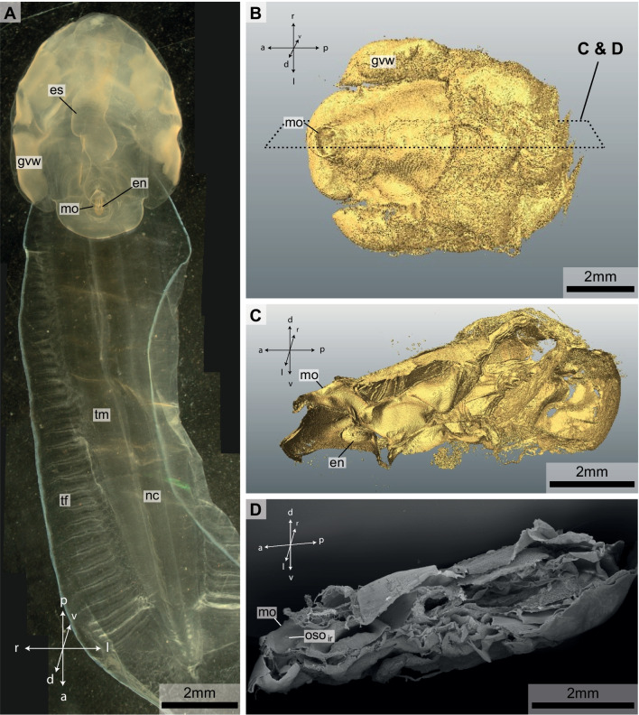
Fig. 1.
A Light micrograph of a specimen of Bathochordaeus stygius. B µCT-isosurface image of the specimen shown in A in dorsal view. Dashed rectangle indicates cutting plane from figures (C and D). C µCT-isosurface image of the trunk of the animal in lateral view from the left side. The left half of the animal was digitally removed along the mid-sagittal plane to show inner structures. D Scanning electron micrograph of the half of the same specimen sectioned along the mid-sagittal plane. Note the mouth opening (mo) to the left and the inner oral sensory organ of the right side (osoir) inside the mouth cavity. In B, C and D orientation is anterior to the left posterior to the right. es—esophagus, en—endostyle, mo—mouth opening, nc—nerve chord, tm—tail muscle, oso—oral sensory organ, gvw—genito-visceral wing, tf—tail fin

