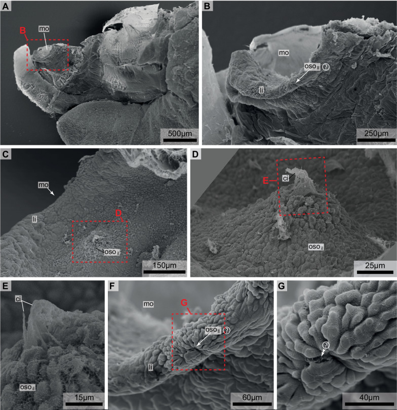
Fig. 2.
Scanning electron micrographs of Bathochordaeus stygius. Specimen cut along the sagittal mid-plane. A Oblique dorsal view anterior to the left. B Oblique dorsal view of mouth opening. C Lateral view of the anterior mouth cavity. Anterior to the upper right of the image. D Interior oral sensory organ of the left side. E Ciliary tuft of the interior oral sensory organ of the left side. F Lateral lip. Note irregular shape of epidermis cells. G Magnification of F depicting possible cilia of the lateral oral sensory organ of the left side (double arrowheads). ci—cilia, li—lip, mo—mouth, osoil—inner oral sensory organ of the left side, osoll—possible lateral oral sensory organ of the left side

