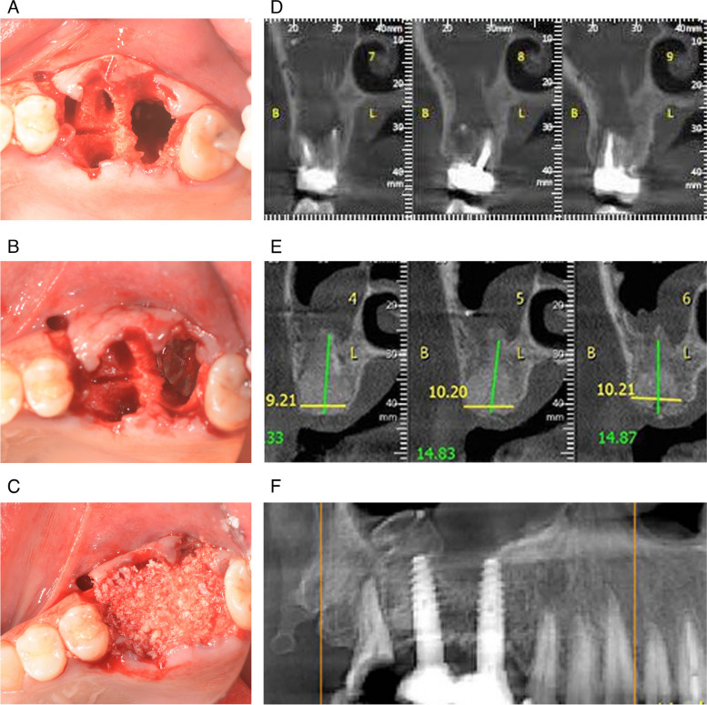
Fig. 2.
A After serial extraction of teeth 16 and 17, oroantral communication at the site of the maxillary second molar was clinically noted and diagnosed. B The magnesium membrane (black arrow) was folded and placed in a way that the communication was closed. C Allograft and xenograft granules were used for augmentation and a second magnesium membrane was placed over the augmentation. D Coronal preoperative CBCT shows that the roots of maxillary second molar were placed directly in the right maxillary sinus. E CBCT four months after graft placement shows that the graft has integrated with the bone and that there was sufficient buccal and palatal bone to place two implants. F Panoramic image 8 months post-operatively shows two integrated implants with no signs of peri-implantitis

