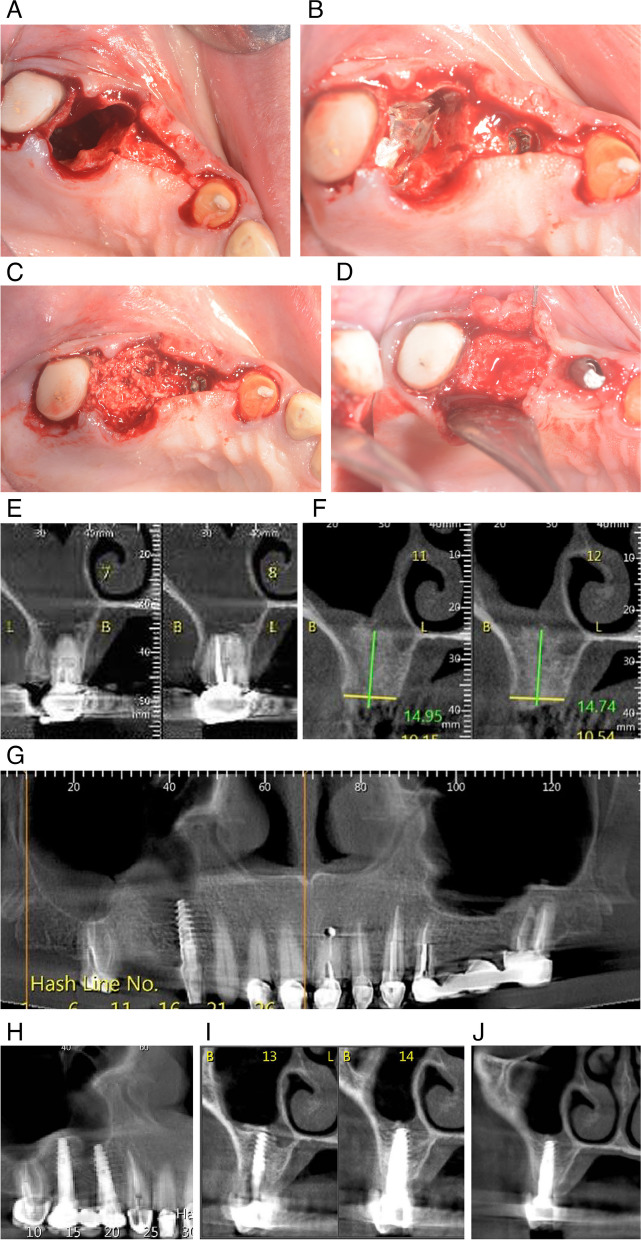
Fig. 3.
A Tooth 16 was extracted, leaving communication between the oral cavity and the maxillary sinus. B The sulcus was opened through a crestal incision and the dental implant was placed at position 14 (blue arrow). Resorbable magnesium membrane was shaped to close the communication at position 16 (black arrow). C The cavity at position 16 was filled with allograft and xenograft granules, the soft tissue was sutured, and a healing cap was placed on the implant (blue arrow). D Five months post-operatively, the implant was stable, with no abnormal movement, and the graft material was integrated with surrounding healthy hard tissue. E Coronal CBCT section taken before extraction shows that there is no alveolar bone between the apical third of tooth 16, which ends in the right maxillary sinus. F CBCT cross-section taken five months after graft placement shows 15 mm of newly formed bone, sufficient for placement of a new fixed restoration. G Panoramic image taken five months after implant placement at position 14. Newly formed bone tissue has formed adjacent to the implant with no signs of peri-implantitis. H The panoramic image was taken four months after implant placement at position 16 in the maxilla. A dental bridge is attached to implants 14 and 16 as well as healthy tissue around the previously placed implants. I, J Coronal CBCT images slices show correct placement of the implant between the buccal and palatal alveolar walls and the presence of new alveolar bone in the apical third of the implant, creating complete separation from the right maxillary sinus

