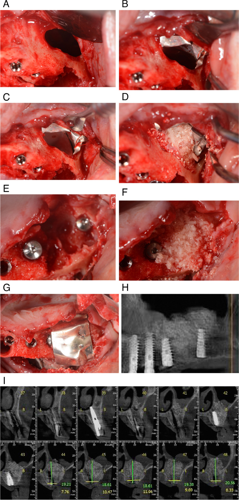
Fig. 4.

A Extraction of tooth 26 resulted in a large loss of bone tissue. A direct approach was used to open the buccal wall to facilitate access to oroantral communication (black arrow). B A magnesium membrane was placed over the buccal window to close the connection between the maxillary sinus and the extraction site (blue arrow). C Second layer of membrane is applied on top of the first layer (blue arrow). D The site was augmented using allograft and xenograft granules. E Four dental implants were immediately placed. F Allograft and xenograft granules were also used to augment the extraction site. G A magnesium membrane was used as a barrier to create space between bone and soft tissue (blue arrow). H Panoramic image four months after placement of four implants at positions 23, 24, 25, and 27 and application of graft material shows stability and formation of new alveolar bone. I Coronal CBCT image shows 18–20.5 mm of alveolar bone between the oral cavity and the floor of the left maxillary sinus
