Fig. 5.
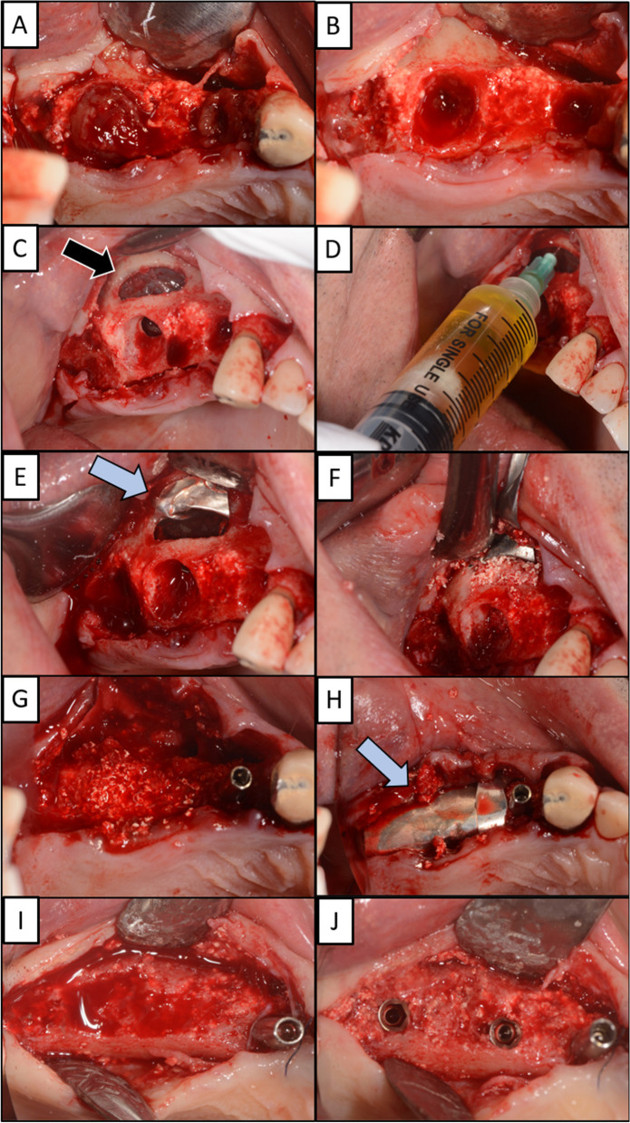
A Extraction of dental implants at position 14 and 16 resulting in large bone defects. B After removal of the additional soft tissue at the extraction site, there is a better view of the oroantral communication. C The window on the buccal wall was surgically opened to gain direct access to the Schneiderian membrane (black arrow). D Aspiration needle was used to aspirate the polyp inside the right maxillary sinus through the Schneiderian membrane. E A magnesium membrane (blue arrow) is placed on Schneiderian membrane to stimulate and form the separation of the oral cavity from the right maxillary sinus. F The buccal window is closed with allograft and xenograft granules. G Immediately after extraction, an implant is placed in position 14, the site of the previous implant, since a greater amount of alveolar bone is preserved here compared to position 16. H A magnesium membrane (blue arrow) is placed as a barrier on extraction site 16 to cover the graft and separate it from the soft tissue. The soft tissue is sutured to achieve primary healing. I Five months later, the magnesium membrane has resorbed and new hard tissue has formed at site 16, which can be used to place new dental implants. J Two dental implants are placed at position 15 and 17

