Fig. 1.

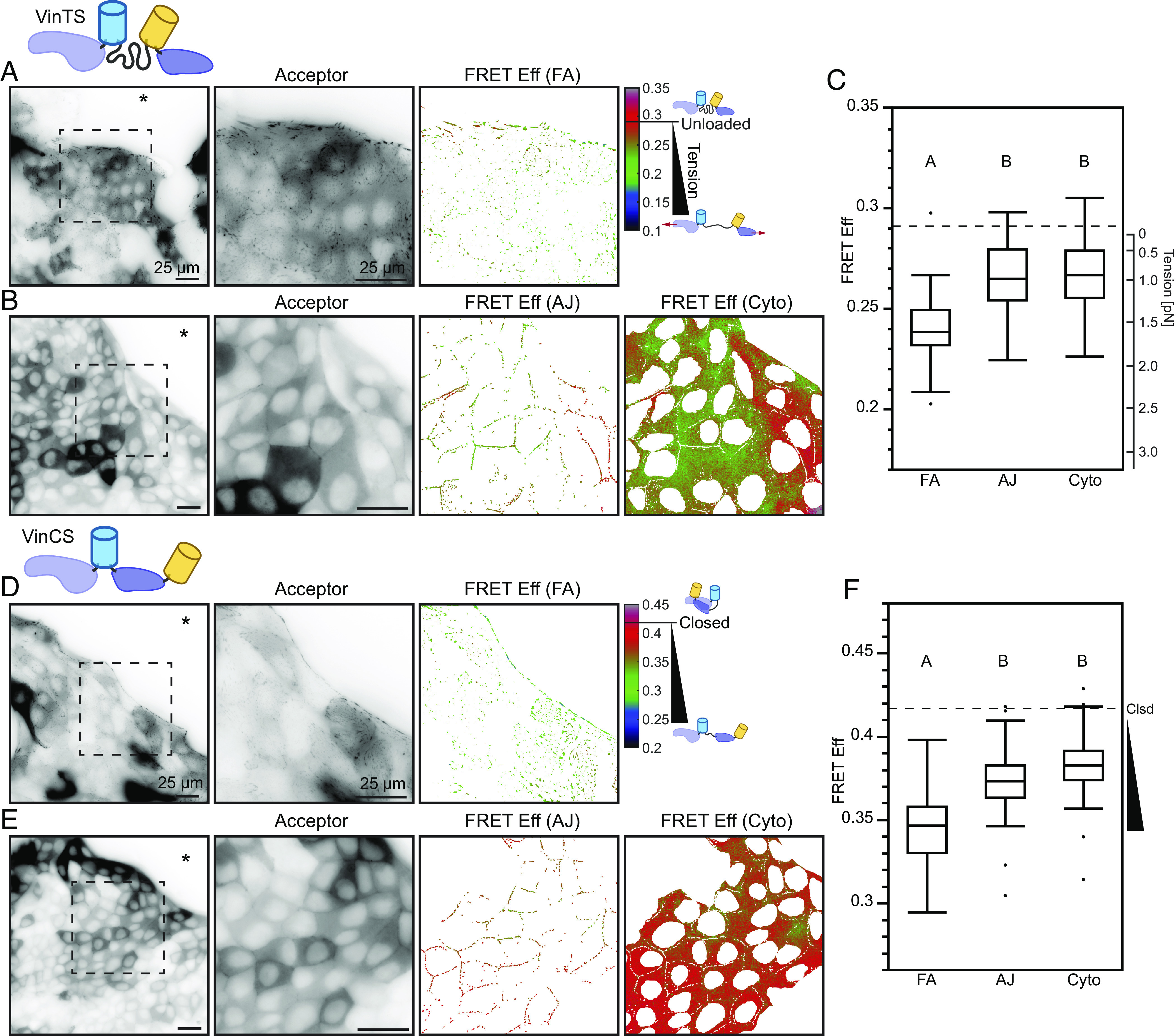
Vinculin is loaded and conformationally open at the edge of collectively migrating cells. Representative images of migrating MDCK II cell monolayers expressing VinTS taken in the basal (A) or apical (B) plane at the monolayer edge with acceptor channel indicating sensor localization followed by zoom-ins of the indicated region for acceptor channel and FRET efficiency in the FA mask (A) or AJ and cytoplasm masks (B). The asterisk indicates free space adjacent to monolayer edge. (C) Box plot of FRET efficiency for VinTS at FAs, AJs, and cytoplasm (n = 43, 34, and 34 images completed over at least 3 independent experiments) with unloaded reference level indicated (dotted line). Representative images of migrating MDCK II cell monolayers expressing VinCS taken in the basal (D) or apical (E) plane at the monolayer edge with acceptor channel indicating sensor localization followed by zoom-ins of the indicated region for acceptor channel and FRET efficiency in the FA mask (D) or AJ and cytoplasm masks (E). (F) Box plot of FRET efficiency for VinCS at FAs, AJs, and cytoplasm (n = 61, 51, and 52 images completed over at least 3 independent experiments) with closed reference level indicated (dotted line). Differences between groups were detected using the Steel–Dwass test. Levels not connected by the same letter are significantly different at P < 0.05. P-values for all comparisons can be found in SI Appendix, Supplementary Note 2 and Table S4 for VinTS and SI Appendix, Supplementary Note 2 and Table S5 for VinCS.
