Fig. 1.

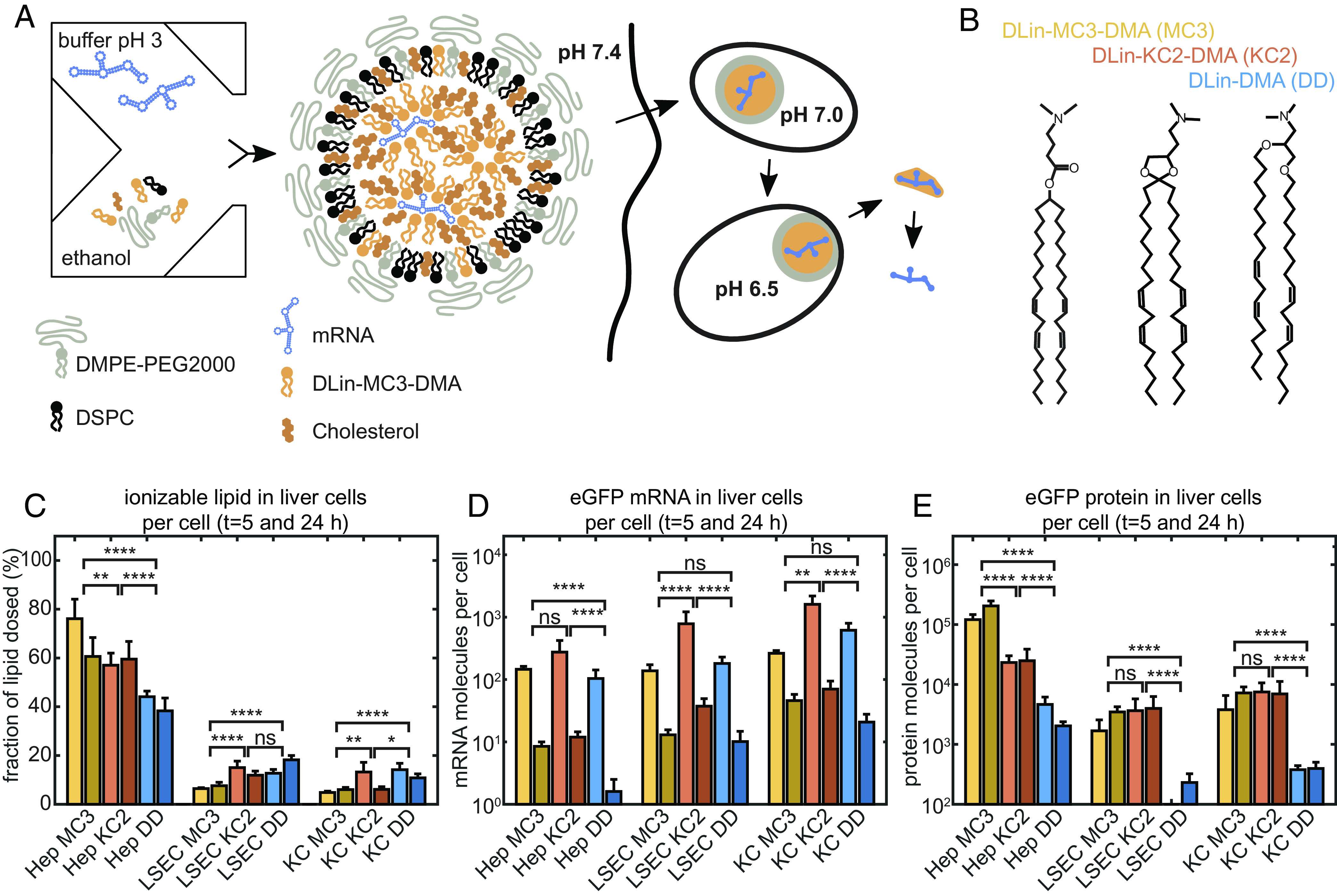
LNP-mediated mRNA delivery and protein expression in vivo. (A) Schematic drawing of production, internalization via endocytosis, and pH-dependent maturation of LNPs preceding endosomal release. mRNA LNP particles exhibit a core-shell structure consisting of a condensed core phase composed of mRNA, CIL, and chol, with an outer shell enriched in DSPC and a PEG corona. (B) The ionizable lipids MC3, KC2, and DD differ in head group area while sharing the same lipid tails and ionizable dimethylamino-group. (C) Mice study of ionizable lipid uptake in liver cells, (D) mRNA uptake in liver cells, and (E) in vivo protein expression per cell type showing same uptake yet unexplained order of protein expression efficacy MC ≥ KC2 > DD. Four mice in each group were used with an mRNA dose of 0.5 mg/kg. Error bars in figures (C–E) are the SEM. Significance levels are based on the Mann–Whitney method.
