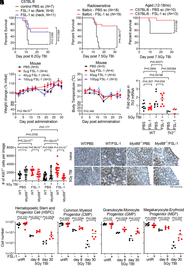
Fig. 1.
FSL-1 effectively mitigated acute radiation-induced lethality in mice by minimizing adverse effects and enhancing proliferation of hematopoietic progenitor cells. (A) C57BL/6 mice were administered FSL-1 subcutaneously (sc) in abdominal flank or scruffed neck 24 h after 8.2 Gy TBI. (B) Balb/c mice were administered FSL-1 (sc) at 24 h after 7.5 Gy TBI. (C) Aged C57BL/6 mice (12 to 18 mo) were given FSL-1 (sc) after 7.5 Gy TBI. Survival distributions in Kaplan–Meier plots and log-rank test P values are shown. Investigating adverse effects, naive mice were given a single FSL-1 sc injection (5 to 100 µg), and changes in weight (D) or body temperature (E) were monitored with mean ± SEM shown from a representative experiment of three replicate studies. Repeated measurements were assessed using linear mixed-effects modeling with P values for treatment to time interaction indicated. (F) qPCR studies were conducted to assess Tlr2 mRNA in bone marrow (BM) cells from WT and Tlr2−/− mice treated with radiation (5 Gy TBI) with (+) and without (−) FSL-1. (G and H) Femur tissue sections prepared at day 8 post treatment were probed with Ki67 antibodies, imaged and proliferating cells were quantified using ImageJ. Scale bar at 100 µm shown on representative images. Pairwise t tests (unpaired data, unequal variance, two-sided) were applied with P values shown and mean indicated by bar. (I) BM hematopoietic stem and progenitor cells (HSPCs), (J) common myeloid progenitors (CMPs), (K) granulocyte-macrophage progenitors (GMPs), and (L) megakaryocyte-erythroid progenitors (MEPs) in unirradiated (unIR) and irradiated mice BM harvested on days 8 and 30 after PBS or FSL-1 sc injections on day 1 were immunophenotyped by flow cytometry. Two-sample t tests were performed for unpaired BM cell data with Welch’s approximation assuming variance between treatment groups. P values are indicated. Each symbol represents an individual mouse (F and I–L) or image of fixed bones (G).

