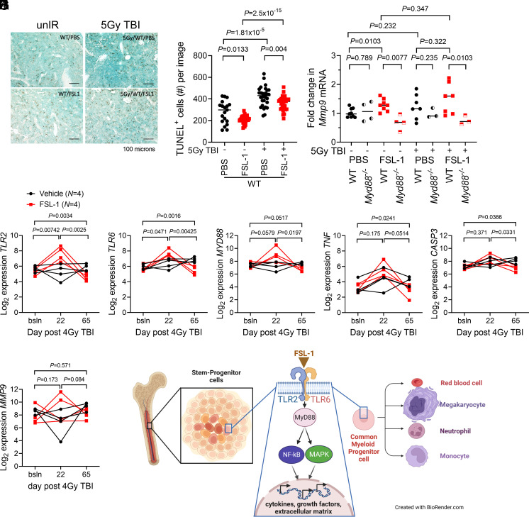
Fig. 4.
FSL-1 stimulated TLR2/TLR6 signaling and activated downstream transcriptional regulation. (A) TUNEL immunohistochemistry of murine femur sections was conducted to study cell death. Femur tissue sections prepared from treated C57BL/6 mice on day 8 after radiation were probed with TUNEL HRP reagent. The scale bar is 100 µm. (B) Quantitation of number (#) of TUNEL+ cells per image was conducted using ImageJ, with each symbol representing an individual femur sample. Statistical significance was determined using pairwise t tests, with P values indicated. (C) Mmp9 mRNA from BM cells harvested at 8 d from FSL-1-treated control and irradiated mice was quantified by qPCR analysis. Each symbol represents an individual mouse with bar indicating the mean. Data (unpaired, unequal variance, two-sided) were evaluated using t tests (unpaired data), with P values indicated. (D–I) Transcript profiling of NHP RNA was conducted on BM biopsies from Vehicle (N = 4) and FSL-1 (N = 4) treated irradiated NHP using NanoString technology. String plots for differentially expressed genes (TLR2, TLR6, MyD88, TNF, CASP3, and MMP9) are shown, where connected lines between symbols represent an individual NHP. The t test (unpaired data) was used to decipher changes in transcripts between FSL-1 and Vehicle treatments at bracketed time points between bsln to d22, d22 to d65 or bsln to d65. (J) A mechanism is proposed whereby FSL-1 binds TLR2/TLR6, resulting in activation of MyD88, NF-κB, and MAPK activities, and culminating in transcriptional regulation of cytokines, growth factors, and extracellular matrix components that promote proliferation of hematopoietic progenitors.

