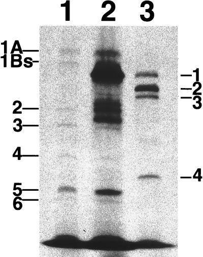
FIG. 1.
Fluorography of PBPs labeled with [benzyl-14C]penicillin after separation by SDS–8% polyacrylamide gel electrophoresis. Lane 1, an E. coli MC1061 transformant having pAW119; lane 2, an E. coli MC1061 transformant having a 4.9-kb genomic fragment of S. aureus NCTC8325; lane 3, S. aureus BB255. Designations of PBPs of E. coli and S. aureus are indicated at the left and right sides of the fluorography, respectively. On each lane, 100 μg of proteins of the membrane fraction was applied.

