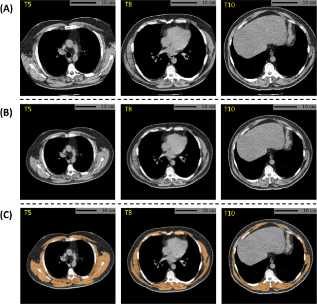
Figure 2:
Example skeletal muscle assessment using lung cancer screening low-dose CT scan of a male participant with a BMI of 35.1 kg/m2 and a smoking history of 41 years (61.5 pack-years). (A) CT axial plane levels corresponding the mid-location of the fifth (T5), eighth (T8), and 10th (T10) vertebral bodies were predicted. (B) The field-of-view of each CT section with body section truncation was extended with missing body section imputation. (C) Cross-sectional regions corresponding to the skeletal muscle were delineated on the field-of-view extended sections. Skeletal muscle attenuation was defined as the averaged radiodensity of muscle regions across the three levels. In this case, the algorithm reported a skeletal muscle attenuation of 17.5 HU, which fell within the lowest quartile (< 27.6 HU) among male participants. BMI=body mass index. HU=Hounsfield unit.

