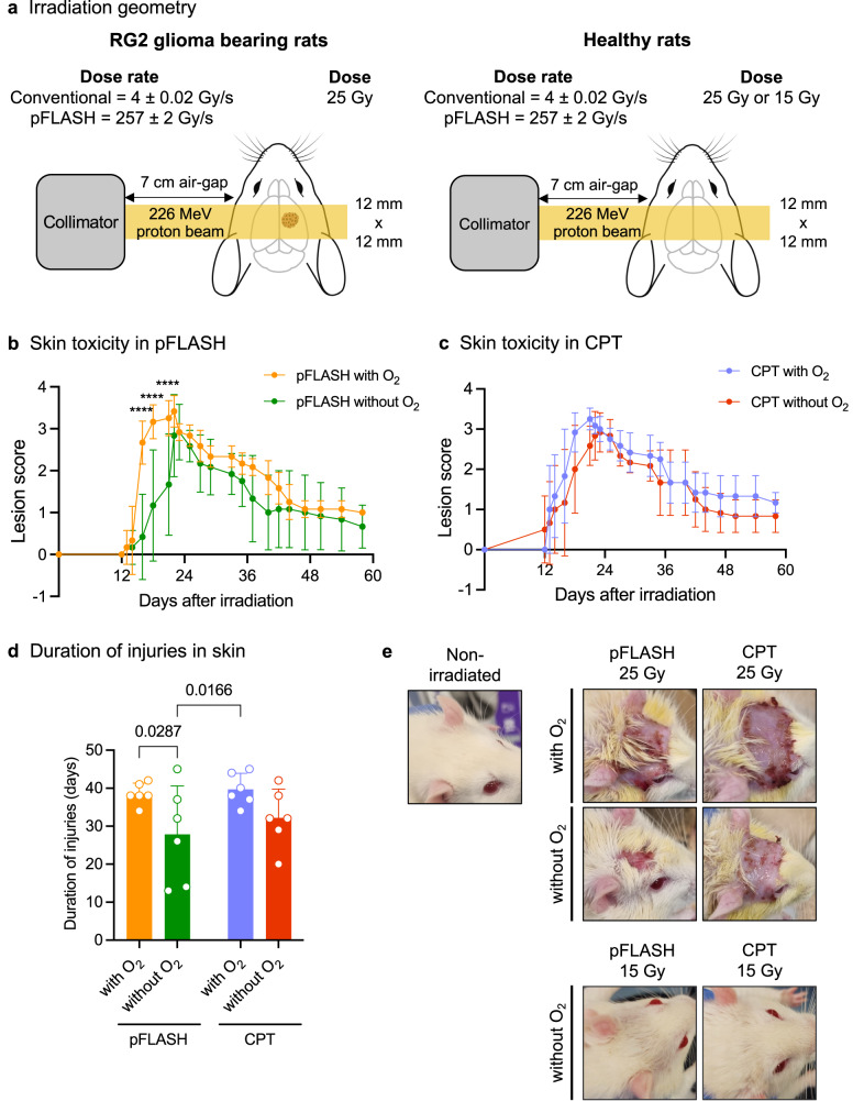
Fig. 1. Impact of oxygen content on the development of radiation dermatitis after pFLASH and CPT delivering 25 Gy.
a Irradiation geometry. All groups received unilateral transmission irradiations using the plateau area of a mono energy 226 MeV proton beam. The original scanned beam was modified into a 12 × 12 mm² collimated scanned beam at the irradiation point using a brass collimator (and 7 cm airgap), with a flatness of §5% at maximum dose level. In all cases, the dose prescription was 25 Gy at 1 cm depth in the brain or in the tumor. b Average lesion score after pFLASH irradiation as a function of oxygen level (n = 6 in each group). c Average lesion score after CPT, as function of oxygen level (n = 6 in each group). d Duration of injuries among the irradiated groups (n = 6 in each group). e Representative images of radiation dermatitis among the groups. Images in the groups receiving 25 Gy (n = 6 in each group) at 22 days post-irradiation (maximal severity), correspond to the mean grade observed on this day (grade 3 for pFLASH with O2, grade 2.5 for pFLASH without O2, grade 3 for CPT with O2, grade 3 for CPT without O2). The rats receiving 15 Gy in CPT or pFLASH did not develop radiation-induced injuries. The data are presented as the mean ± standard deviation (SD).

