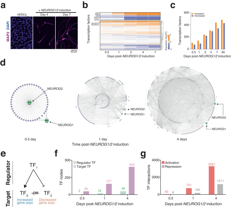
Fig. 1. Integrating gene expression and chromatin accessibility to assemble transcription factor (TF) networks during the earliest stages of human neuron differentiation.
a Representative immunofluorescence images of human embryonic stem cells and NEUROG1/2-induced human neurons stained with the neuronal marker microtubule-associated protein 2 (MAP2, purple) and the nuclear stain DAPI (blue). b Fold-change in the expression of all TFs over 5 timepoints during differentiation (n = 4 biological replicates at each time point). c Number of TFs with altered expression in developing iNs. d Network diagrams of TF at different timepoints after NEUROG1/2 induction. Lines indicate a regulatory interaction between TF pairs. e Regulator TFs can act to either activate or repress their target TFs. f Number of regulators and target TFs at different timepoints after NEUROG1/2 induction. g The number of activating and repressing TF interactions at different timepoints after NEUROG1/2 induction.

