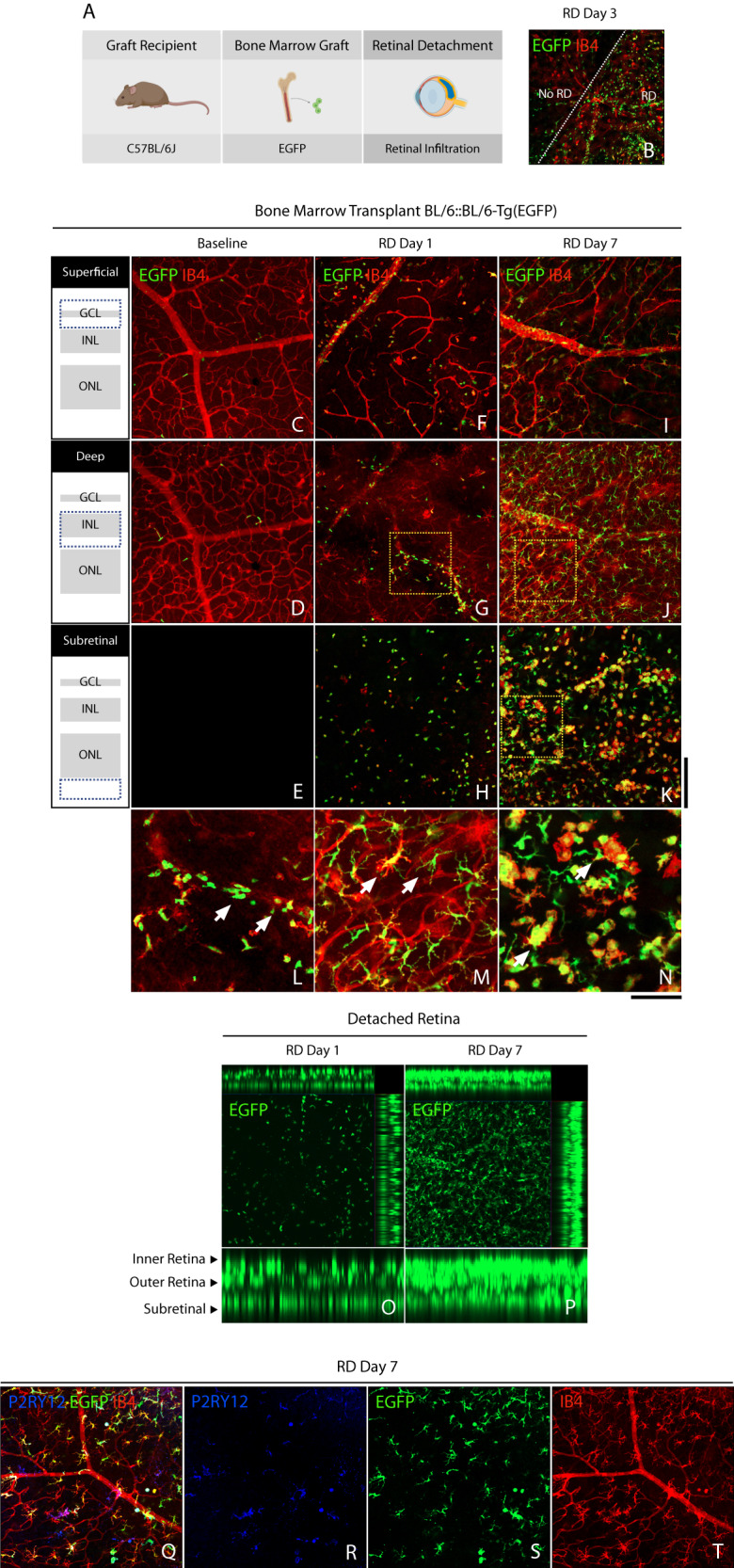
Fig. 2. Peripheral myeloid cell infiltration in retinal detachment.

A Outline of recipients, bone marrow transplantation, and experimental retinal detachment model (Created with BioRender.com). B Representative image of retinal wholemounts of chimeric mice at day 3 following retinal detachment showing the attached (No RD) and detached retina (RD) separated by a white dotted line. C–E Representative confocal image of chimeric mice at baseline. F–H Detached retina at day 1, with moderate peripheral EGFP+ BM-derived infiltration of the retina and subretinal space. I–K Detached retina at day 7, with significant EGFP+ infiltration of the retina. L Magnification of image G (dotted box), showing early retinal infiltration by peripheral EGFP+ cells of ameboid shape (arrows). M Magnification of image (J) (dotted box), showing substantial retinal infiltration by EGFP+ cells with ramified phenotype (arrows). N Magnification of image (K) (dotted box), showing mixed ameboid and ramified morphology on EGFP+ cells in the subretinal space. O and P Z-projection of confocal images showing peripheral EGFP+ cell infiltration in the detached retina and subretinal space on days 1 and 7. Q–T Scarce P2RY12+ retinal microglia surrounded by ubiquitous myeloid-derived EGFP+ cells in the detached retina. Images are representative of ≥3 experiments. Scale bar 100 µm (A–I).
